Paper Links
Full Paper (Open-Access)
Abstract
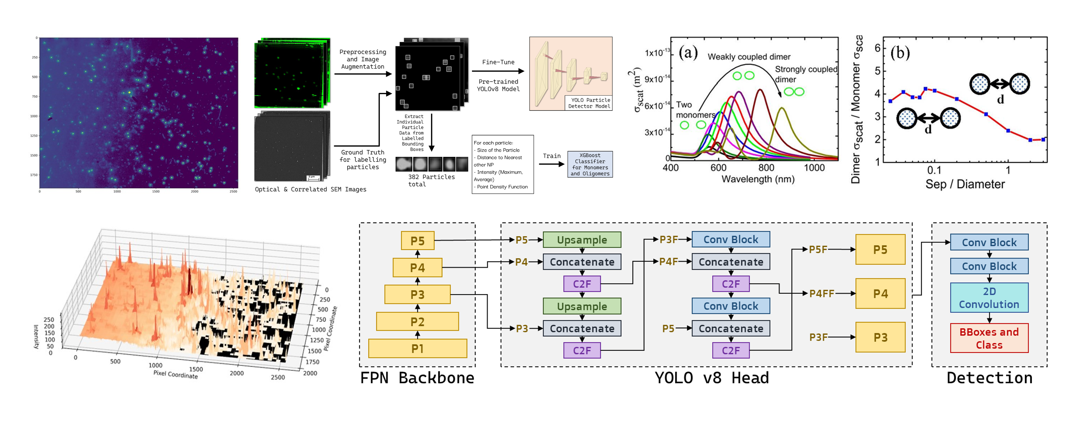
Nanoparticles embedded in polymer matrices play a critical role in enhancing the properties and functionalities of composite materials. Detecting and quantifying nanoparticles from optical images (fixed samples - in vitro imaging) is crucial for understanding their distribution, aggregation, and interactions, which can lead to advancements in nanotechnology, materials science, and biomedical research. In this paper, we propose an ensembled deep learning approach for automatic nanoparticle detection and oligomerization quantification in a polymer matrix for optical images. The majority of prior studies of nanoparticle identification and categorization of fixed samples was based on scanning electron microscope (SEM) or transmission electron microscope (TEM) images which are destructive to biological imaging. However, the proposed study is based on optical images, which are susceptible to noise, low contrast, anisotropic shape, overlapping of the point spread function, plasmon coupling, and resolution limitations. In this study, we fine tune a deep neural network architecture, YOLOv8, on a carefully annotated dataset of correlated optical and SEM images of 80 nm gold nanospheres (AuNS) of varying oligomerization states. The resultant model features a weighted average accuracy of 80.7% for quantification of AuNSs and determining their oligomeric state, far surpassing the capabilities of existing manual image processing methods. We also demonstrate its speed and effectiveness in nanoparticle detection and oligomerization within the polymer matrix through tests on high density uncorrelated optical images. The optical image-based quantification technique will be useful for (live samples - for in vivo imaging) analyzing nanoparticle uptake, oligomerization state and aggregation kinetics in live cells, identifying stoichiometry of membrane protein and its interactions, nanoparticle cell interaction, cell signaling imaging, and drug delivery.
Code
Dataset
Please contact the authors for access to the dataset.
Credits
Shadab Hafiz Choudhury 1 and Dr. Abu Mohsin 1 2
1: Department of Electrical and Electronic Engineering, Brac University
2: Corresponding Author
Both authors contributed equally to this work.
Citation
@article{mohsin_quantifying_2025,
	title = {Quantifying {Monomer}–{Dimer} {Distribution} of {Nanoparticles} from {Uncorrelated} {Optical} {Images} {Using} {Deep} {Learning}},
	url = {https://doi.org/10.1021/acsomega.4c07914},
	doi = {10.1021/acsomega.4c07914},
	urldate = {2025-01-07},
	journal = {ACS Omega},
	author = {Mohsin, Abu S. M. and Choudhury, Shadab H.},
	month = jan,
	year = {2025},
	note = {Publisher: American Chemical Society},
}